Object Record
Images

Metadata
Object Name |
Access Set |
Object ID |
2008.0144b |
Description |
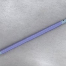
Access Set, 2008 This set consists of multiple instruments. A long thin needle with a mandrel inside is pushed into the hollow system of the kidney after it has been filled from below with contrast so that we can see it easily. Next the mandrel is removed; the needle is left in place and a thin wire is inserted through the needle. This guidewire has a flexible tip, which allows it to move through the needle, through the kidney, into the ureter. The needle is withdrawn and a stiff sheath is inserted over the guidewire. Multiple dilators (the light blue tubes with scale on side) are then inserted until we get to the largest size needed. We then slip over this the dark blue sheath which goes into the kidney; the guidewire is left in place. The dilator is removed, leaving the sheath in place and through this we then insert the ultrasound instruments. Usually we place a second wire which runs outside the sheath to maintain access to the kidney should one of the instruments slip during the procedure. Once the stone is completely destroyed, we can flush out the pieces. When all pieces have been removed either by suction or by removing larger pieces with a grasper, we remove the plastic sheath and place a catheter from the skin into the kidney. 2008.0144 Donated by Boston Scientific |
Collection |
Boston Scientific |
Date |
2008 |
Artist/Designer |
Boston Scientific |
Source |
Boston Scientific |
